

What is patellofemoral chondropathy?
Patellofemoral cartilage anatomy
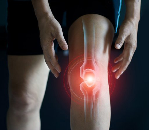
Patellofemoral chondropathy, sometimes called chondromalacia, refers to actual, observed damage to the cartilage covering the back of the kneecap and the femoral trochlea it glides over. It differs from patellofemoral pain syndrome (or patellar syndrome), which is a functional disorder without necessarily any visible abnormality on imaging: chondropathy, by contrast, is a structural cartilage lesion confirmed by an examination.
Why does this cartilage get damaged?
Cartilage damage results from repeated mechanical overload of the patellofemoral joint over time, often linked to chronic poor kneecap tracking, intensive sporting activity with many weight-bearing loads on a flexed knee, or a history of direct trauma to the kneecap (a fall, an impact).[1]
Symptoms and diagnosis of patellofemoral chondropathy
Typical pain and its triggers
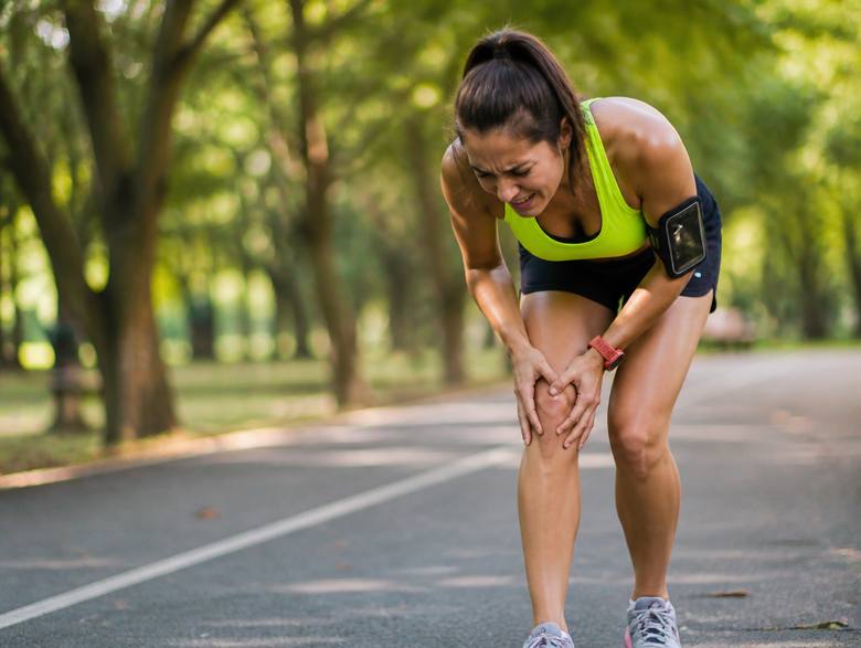
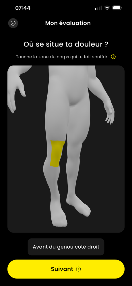
The pain sits at the front of the knee, spread around or behind the kneecap, and often comes with a noticeable (sometimes audible) grinding or grating sensation during knee flexion-extension. It’s typically triggered by sitting for a long time with bent knees (the “cinema sign”), going down stairs, squatting, and deep squats.[1] In more marked cases, mild, non-red swelling can appear after a heavy session.
Differential diagnosis
Several types of front-of-knee pain can be mistaken for chondropathy, which is why a precise assessment matters before starting a suitable programme:
- Patellofemoral pain syndrome, a functional disorder without necessarily any structural abnormality on imaging
- Patellar tendinopathy, with pinpoint pain on the tendon just below the tip of the kneecap, triggered mainly by jumping
- An associated meniscal or ligament injury, if there’s a history of knee trauma
A precise description of your symptoms (location, grinding, triggers) is what first guides the diagnosis; imaging is only useful if there’s doubt or if the pain resists rehab.
Causes and risk factors
Cartilage damage almost always results from repeated mechanical overload of the patellofemoral joint over time, rather than a single isolated event.
Risk factors
- Chronic poor kneecap tracking, with the kneecap poorly centred in the trochlea
- Weak quadriceps, particularly the inner portion (vastus medialis), and weak hip muscles that leave the kneecap poorly stabilised[1]
- A history of direct trauma to the kneecap (a fall, an impact)
- Being overweight, which increases the strain transmitted with every knee bend
- Intensive, prolonged sporting activity with many weight-bearing loads on a flexed knee
- Getting older, as cartilage gradually tolerates repeated strain less well over time
Which sports contribute to patellofemoral chondropathy?
All sports that intensely and repeatedly load the flexed knee carry a risk of this cartilage wear:
- High-volume running over many years
- Intensive cycling
- Volleyball and basketball (repeated jumping)
- Weight training with repeated deep squats
- Skiing
- Sports with a history of direct trauma to the kneecap
Treating patellofemoral chondropathy in rehab with a sports physio
The goal of treatment is to reduce mechanical strain on the damaged cartilage area while strengthening the muscles that stabilise the kneecap, to slow progression and significantly improve everyday and sporting comfort.
- 1Hip and knee mobility
- 2Strengthening the quadriceps (vastus medialis)
- 3Strengthening the hip muscles (gluteus medius)
- 4Correcting weight-bearing and the kinetic chain
- 5Eccentric strengthening
- 6Working on the posterior chain
- 7Progressive reintroduction of running
- 8Light plyometrics
- 9Progressive return to your sport
- 10Return to competition
“Cartilage doesn’t regenerate like a muscle, but a well-strengthened, well-loaded joint stays a joint that works.”
Strengthening and load management, the cornerstones of treatment
Strengthening the quadriceps and hip stabilising muscles better distributes strain on the patellofemoral joint, reducing direct pressure on the damaged cartilage area with each knee bend.
In brief
- Strengthening the quadriceps, particularly its inner portion
- Strengthening the hip to stabilise the pelvis
- Suitably managed sporting load
- Gradual return: mobility, then load, then sport
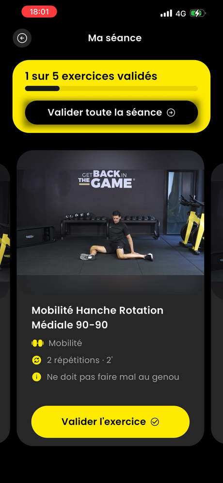
Ready to take your patellofemoral chondropathy seriously?
Start the HOPP programme designed by sports physios.
Should you rest or keep playing sport?
Total rest is generally not the best option for established chondropathy: well-dosed muscle strengthening, combined with suitably managed load, best preserves joint function over the long term.
Think you might have a different knee injury?
Take your free test in 3 minutes flat.
When should you see a specialist for patellofemoral chondropathy?
A medical opinion is recommended to confirm what’s going on with a clinical examination and suitable imaging, particularly in the case of recurrent swelling, mechanical locking, or pain that worsens despite rehab.
How long does it take to recover from patellofemoral chondropathy?
Cartilage doesn’t repair itself like a muscle: there’s no “healing” timeframe as such. What changes quickly is your pain and your day-to-day function: most athletes who follow a strengthening and load-management programme feel a clear improvement within 6 to 12 weeks, with a gradual return to sport from the first few weeks if pain allows.
What affects this timeframe
- The grade of cartilage damage (mild to severe)
- How long the symptoms had been present before treatment started
- How consistently you keep up muscle strengthening
- Following load management properly (the 10% rule)
More severe damage, or damage neglected for several months, can require several months of regular rehab before a confident return to competition. The key point: progression matters more than speed, returning too quickly is the leading cause of relapse.
Preventing recurrence
Even once stabilised, chondropathy stays sensitive to a poorly dosed return to activity: several measures help limit the risk of recurrence once the pain is under control.
- Keep up regular strengthening of the inner quadriceps and hip over the long term, even once the pain is gone
- Stick to the 10% rule when increasing training volume or intensity week to week
- Temporarily adapt risky movements (deep squats, sitting for a long time with bent knees) during periods of heavy load
- Watch for early signs (more grinding, swelling) and adjust load before the pain settles in for good
- Avoid jumping back into sport abruptly after a long break
The HOPP care programmes to treat your chondropathy and get back to sport
This chondropathy requires well-dosed strengthening and suitably managed load. HOPP offers you a precise exercise programme, allowing you to get back to your activities as quickly as possible.
The programme runs from 3 to 9 weeks depending on the injury and ends with a reconditioning phase allowing a return to competition in the best possible conditions. Each programme is designed by the HOPP team, made up of two sports physiotherapists who are experts in assessing and treating athletes. You’ll also find theoretical content throughout your progression, so you master every aspect of your recovery and can adjust your treatment independently. If needed, our team of physiotherapists is on hand to support you.
Essential
FROM INJURY TO
BACK TO YOUR ACTIVITIES
59€
One-time payment
“AT YOUR OWN PACE WITH
DAILY ADAPTATION”
- 100% of the treatment programme
- Daily personalisation
- Flexibility / Mobility
- Strengthening
- Proprioception
- Progress tracking
- Return-to-running programme
- Theoretical content included
Performance
TO GET BACK TO SPORT
IN THE BEST CONDITIONS
79€
One-time payment
“GET BACK TO YOUR LEVEL
WITHOUT RISK OF RELAPSE”
- 100% of the treatment programme
- Daily personalisation
- Flexibility / Mobility
- Strengthening
- Proprioception
- Progress tracking
- Return-to-running programme
- Theoretical content included
- + 4-week return-to-play plan with 6 difficulty levels
Expert
PERSONALISED VIDEO-CALL
SUPPORT, ANY TIME
149€
One-time payment
“A PHYSIO
WHENEVER YOU NEED”
- 100% of the treatment programme
- Daily personalisation
- Flexibility / Mobility, Strengthening, Proprioception
- Progress tracking
- Return-to-running programme
- Theoretical content included
- + 4-week return-to-play plan with 6 difficulty levels
- + 2 video calls with an expert sports physio / return check-up
A programme that adapts to you every day
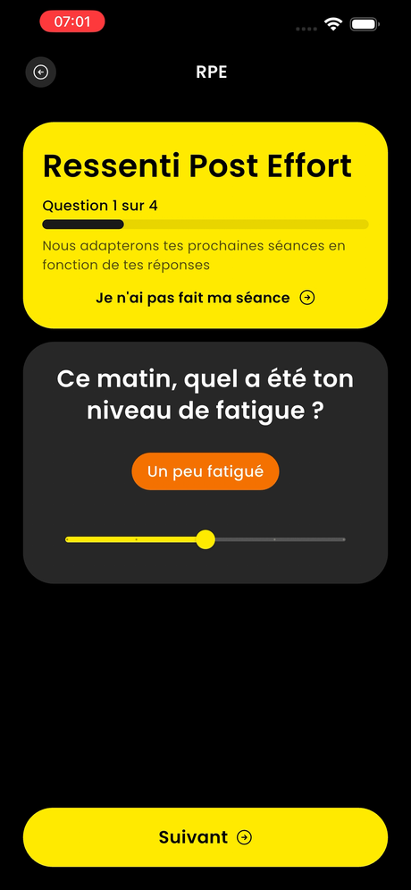
Every day, how you feel (RPE) is factored in to automatically adjust the rest of your programme: more load if all is well, a bit less if your knee is complaining. This daily personalisation is what makes the difference compared to a fixed, paper-based programme.

Ready to start your programme?
Join the HOPP support programme designed by sports physios.
F.A.Q
Does the HOPP programme replace a medical consultation?
No. HOPP is a support and rehab tool designed by sports physiotherapists, but it doesn’t replace a doctor’s opinion. If in doubt, or if the pain persists, see a health professional.
Is the programme suited to all sporting levels?
Yes. Every programme is personalised to your sport, your level and how you feel each day, whether you’re a recreational athlete or preparing for competition.
What’s the difference with patellofemoral pain syndrome?
Patellofemoral pain syndrome is a functional disorder without necessarily any structural abnormality, while chondropathy refers to actual cartilage damage found on examination. The two can coexist.
Should I stop sport with this chondropathy?
Not necessarily. Suitable, well-dosed activity, combined with regular muscle strengthening, is generally more beneficial than stopping completely.
Will this condition get worse?
Suitably managed load and regular strengthening can often slow the progression of symptoms and preserve function in the long run.
Can patellofemoral chondropathy progress to osteoarthritis?
Neglected chondropathy, combined with poorly managed load over several years, can encourage more pronounced cartilage wear. That’s why early treatment and regular strengthening are recommended to protect the joint in the long run.
Do I need an MRI to confirm the diagnosis?
Not systematically. A precise description of your symptoms (location, a grinding sensation, triggers) is often enough to guide the diagnosis. Imaging is mainly useful if there’s doubt, or if pain doesn’t improve despite well-managed rehab.